Heart Anatomy Drawing
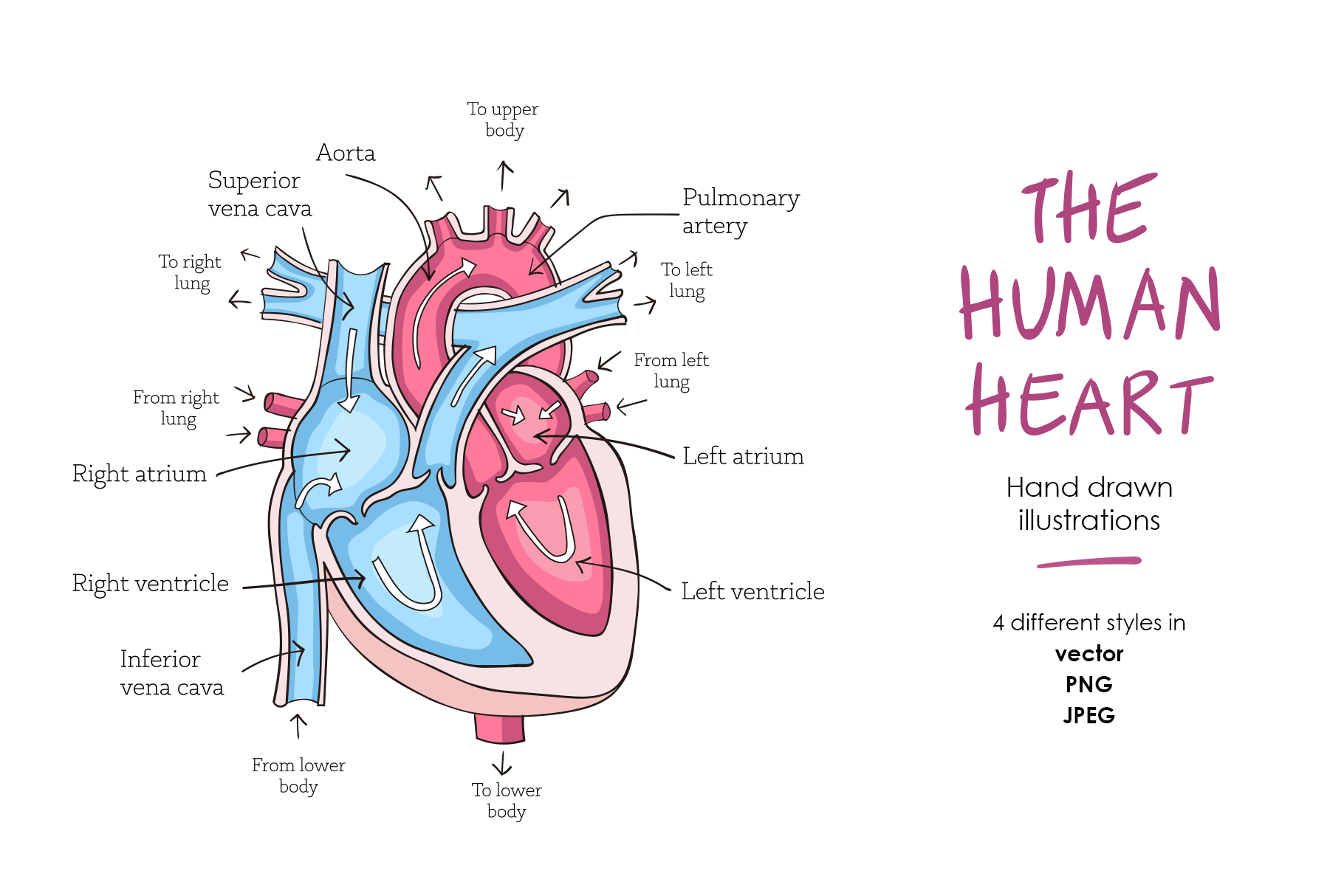
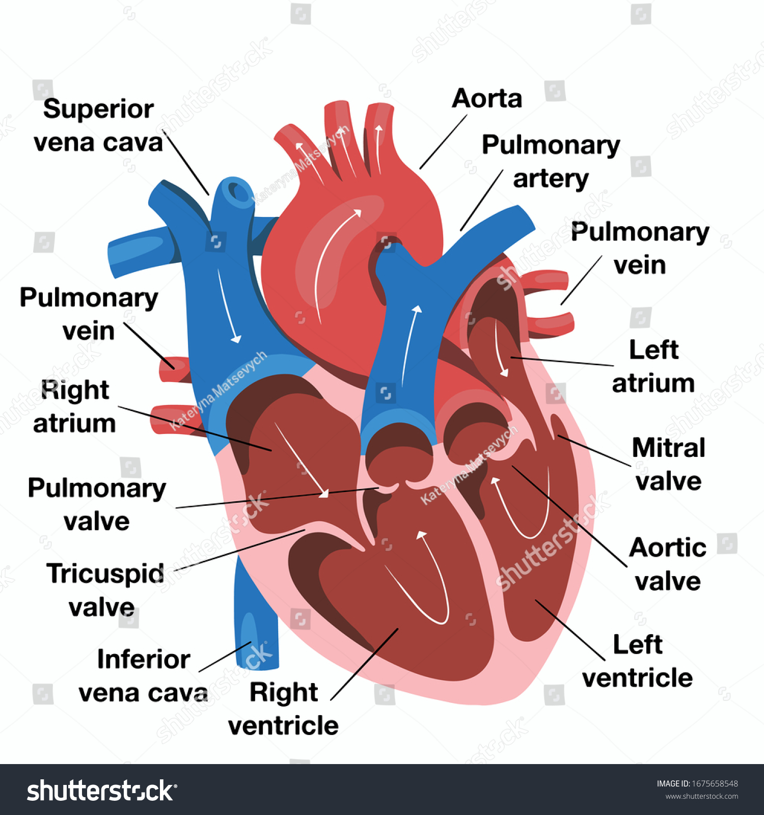
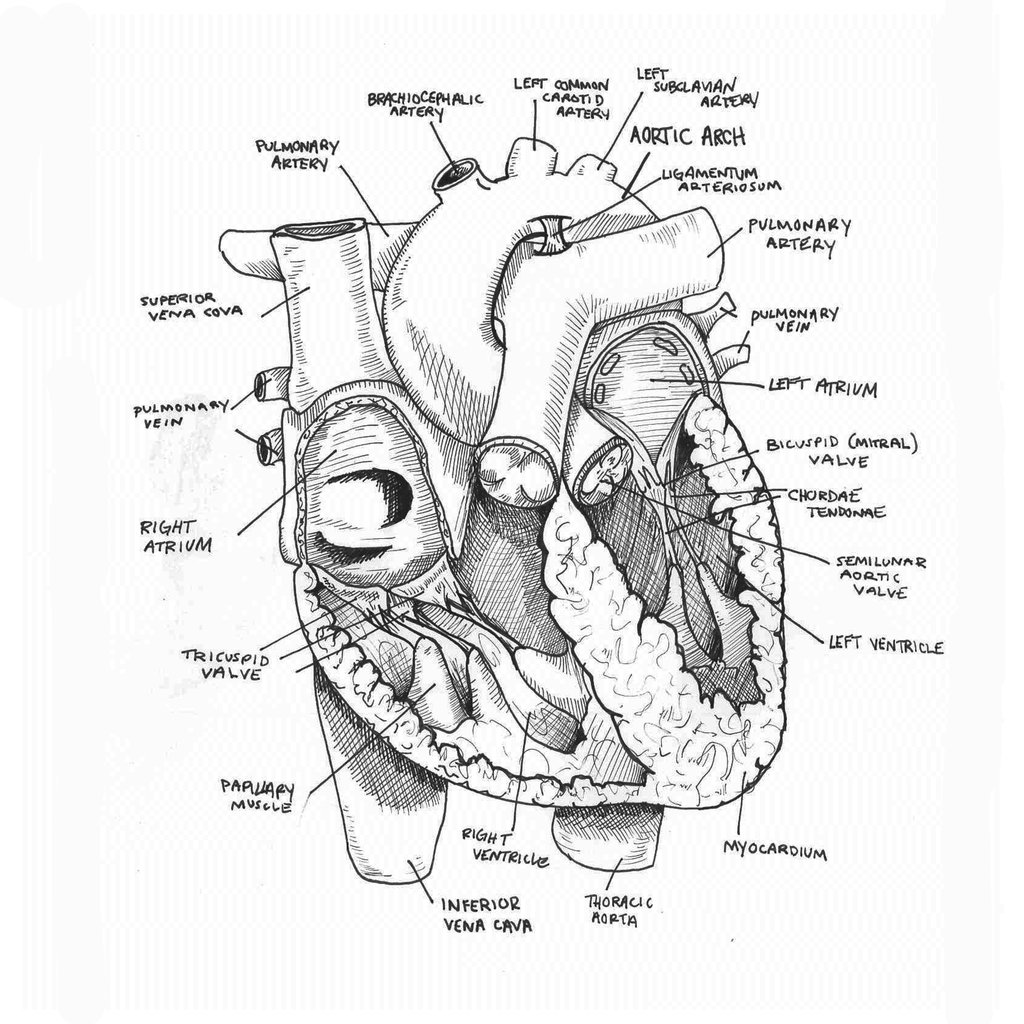
Heart Anatomy Drawing - Web the videos and images on the atlas of human cardiac anatomy are free to download and use for educational purposes. In return, we request that you maintain the university of minnesota/© medtronic watermark on the video/image, and include this citation: Web to draw the internal structure of the heart, start by sketching the 2 pulmonary veins to the lower left of the aorta and the bottom of the inferior vena cava slightly to the right of that. Web to draw an anatomical heart realistically, pay attention to the proportions and positioning of the different parts of the heart, as well as their texture and color. Two atria and two ventricles. Less searching, more finding with getty images. Web explore authentic heart anatomy drawing stock illustrations & vectors for your project or campaign. Web the heart is located in the thoracic cavity medial to the lungs and posterior to the sternum. Web heart, organ that serves as a pump to circulate the blood. The bottom tip of the heart, known as its apex, is turned to the left, so that about 2/3 of the heart is located on the body's left side with the other 1/3 on right. It consists of four main chambers: It is divided into the left and right sides by a muscular wall called the septum. In this lecture, dr mike shows the two best ways to draw. From the openstax anatomy and physiology book. Web heart (right lateral view) the heart is a muscular organ that pumps blood around the body by circulating it through the circulatory/vascular system. In return, we request that you maintain the university of minnesota/© medtronic watermark on the video/image, and include this citation: You can start learning the anatomy of the heart with the following quiz. Web explore authentic heart anatomy drawing stock illustrations & vectors for your project or campaign. The bottom tip of the heart, known as its apex, is turned to the left, so that about 2/3 of the heart is located on the body's left side with the other 1/3 on right. It’s an example of a drawing that can give you a basic idea of what a human heart sketch should be look like. This outlines the lower chamber of the heart, which includes both the left and right ventricles. Understanding its basic anatomy is crucial to understanding how it functions. Begin by sketching a rounded, lumpy, irregular figure. Two atria and two ventricles. It consists of four main chambers: Web these anatomical heart medical illustrations are highly detailed drawings that blend art with science. Let you see a structure from multiple perspectives, for extra clarity; Less searching, more finding with getty images. The user can show or hide the anatomical labels which provide a useful tool to create illustrations perfectly adapted for teaching. Two atria and two ventricles. Let you see a structure from multiple perspectives, for extra clarity; This outlines the lower chamber of the heart, which includes both the left and right ventricles. Web the intricate anatomy of the heart can be challenging to grasp, and so i hope you find this tool to be helpful in visualizing the cardiac system. The user can show or. Some shading quickly helps to. Web challenge your ability to connect anatomy with clinical practice; Next you will draw the aortic arch. Then, fill in the base of the heart with the right and left ventricles and the right and left atriums. Sketch out a basic outline of the heart, using our tutorial as a guide. Web explore authentic heart anatomy drawing stock illustrations & vectors for your project or campaign. Some shading quickly helps to. We will then proceed to shape the heart, slowly refining it with our pencils into a. Sketch out a basic outline of the heart, using our tutorial as a guide. Less searching, more finding with getty images. Web explore authentic heart anatomy drawing stock illustrations & vectors for your project or campaign. The heart is a muscular pumping organ located medial to the lungs along the body's midline in the thoracic region. Web the heart is located in the thoracic cavity medial to the lungs and posterior to the sternum. It consists of four main chambers: Two. Web explore authentic heart anatomy drawing stock illustrations & vectors for your project or campaign. The bottom tip of the heart, known as its apex, is turned to the left, so that about 2/3 of the heart is located on the body's left side with the other 1/3 on right. Next you will draw the aortic arch. Understanding its basic. Web your heart sure does work hard, but that doesn’t mean you have to work hard to draw it! Atlas of human cardiac anatomy, university of minnesota/© medtronic (www.vhlab.umn.edu/atlas) Web to draw an anatomical heart realistically, pay attention to the proportions and positioning of the different parts of the heart, as well as their texture and color. Web this interactive. Web these anatomical heart medical illustrations are highly detailed drawings that blend art with science. Begin by sketching a rounded, lumpy, irregular figure. You can start learning the anatomy of the heart with the following quiz. Web the heart is located in the thoracic cavity medial to the lungs and posterior to the sternum. Web the intricate anatomy of the. 48k views 1 year ago cardiovascular system. It consists of four main chambers: Web drawings of the surface anatomy of the normal heart, anterior and posterior, with english labels. Web to draw the internal structure of the heart, start by sketching the 2 pulmonary veins to the lower left of the aorta and the bottom of the inferior vena cava. 48k views 1 year ago cardiovascular system. Less searching, more finding with getty images. It is divided into the left and right sides by a muscular wall called the septum. On its superior end, the base of the heart is attached to the aorta, pulmonary arteries and veins, and the vena cava. Let you see a structure from multiple perspectives, for extra clarity; Included below are a magnificent color heart illustration, along with four monotype prints, which are possibly woodcuts, engravings, or lithographs. Plus, you may just learn something new along the way. Web the videos and images on the atlas of human cardiac anatomy are free to download and use for educational purposes. Web this interactive atlas of human heart anatomy is based on medical illustrations and cadaver photography. Web dr matt & dr mike. Then, fill in the base of the heart with the right and left ventricles and the right and left atriums. Web these anatomical heart medical illustrations are highly detailed drawings that blend art with science. In this lecture, dr mike shows the two best ways to draw. Web function and anatomy of the heart made easy using labeled diagrams of cardiac structures and blood flow through the atria, ventricles, valves, aorta, pulmonary arteries veins, superior inferior vena cava, and chambers. Web to draw the internal structure of the heart, start by sketching the 2 pulmonary veins to the lower left of the aorta and the bottom of the inferior vena cava slightly to the right of that. Web your heart sure does work hard, but that doesn’t mean you have to work hard to draw it!
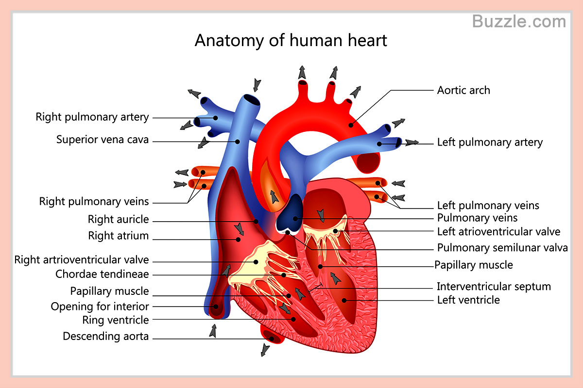
External Structure Of Heart Anatomy Diagram

Heart Anatomy chambers, valves and vessels Anatomy & Physiology
Human heart anatomy (274491) Illustrations Design Bundles
Labeled Drawing Of The Heart at GetDrawings Free download

How to Draw the Internal Structure of the Heart (with Pictures)

Human heart anatomy. Vector diagram in 2021 Heart anatomy, Human

How to Draw the Internal Structure of the Heart 13 Steps
Hand Drawn Illustration Human Heart Anatomy Stock Vector (Royalty Free
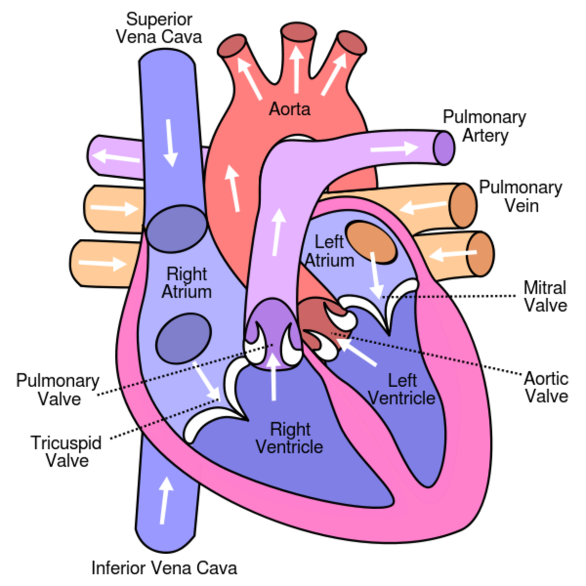
Learn About the Heart and Circulatory System for Kids hubpages
Anatomical Drawing Heart at GetDrawings Free download
The User Can Show Or Hide The Anatomical Labels Which Provide A Useful Tool To Create Illustrations Perfectly Adapted For Teaching.
Some Shading Quickly Helps To.
Next You Will Draw The Aortic Arch.
This Outlines The Lower Chamber Of The Heart, Which Includes Both The Left And Right Ventricles.
Related Post: