Rough Er Drawing
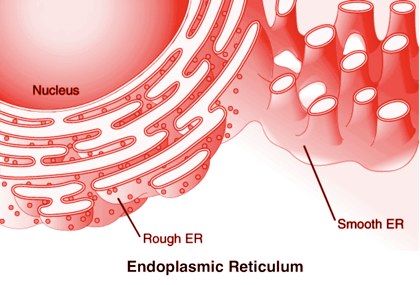
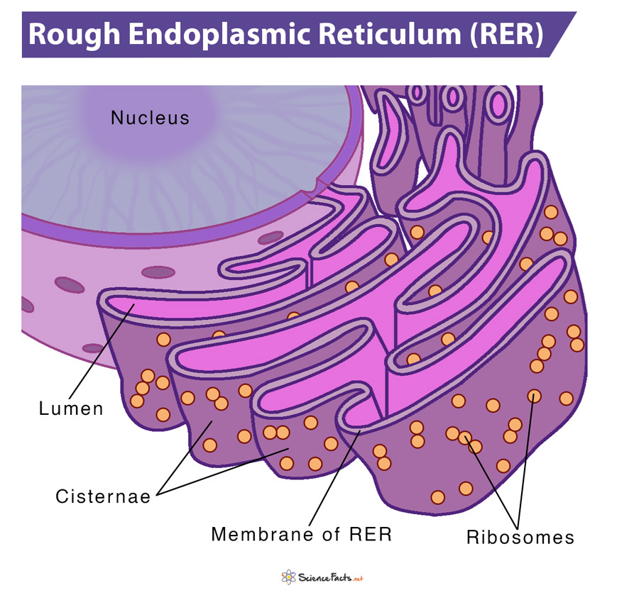
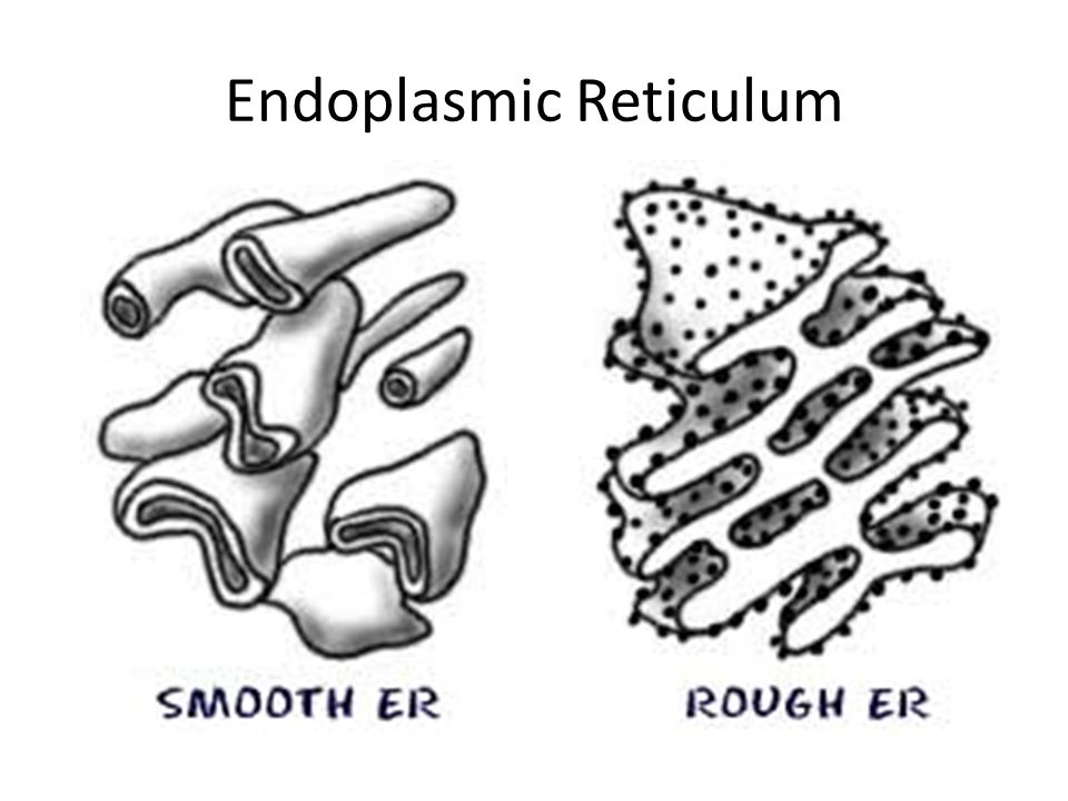
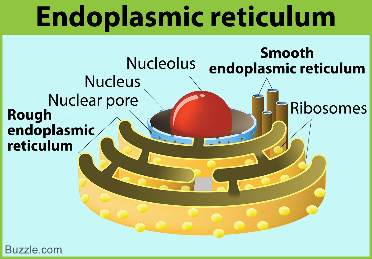
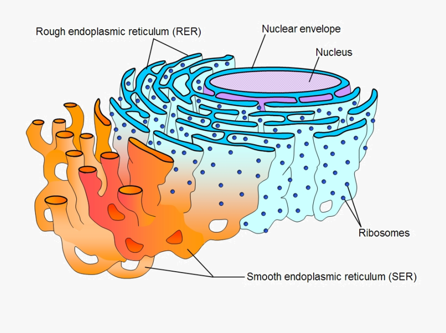
Rough Er Drawing - Web in this video i will show you how can you draw a very important biology diagram endoplasmic reticulum. Web how to draw rough endoplasmic reticulum step by step diagram for class 11th student in the easy way after watching this video. Web hello friends, this is my youtube channel and in this channel i used to share videos of different diagrams in easy way and step by step tutorials. Structure diagram drawing of rough and smooth endoplasmic. Web the rough er is covered with ribosomes where the information carried in messenger rna molecules is translated into proteins. Web what is the endoplasmic reticulum? Web how to draw endoplasmic reticulum. As these ribosomes make proteins, they feed the. Web after watching this video completely you will understand how to draw endoplasmic reticulum. The endoplasmic reticulum transpires in two forms: Web after watching this video completely you will understand how to draw endoplasmic reticulum. Web the er can be classified in two functionally distinct forms: The rough endoplasmic reticulum (rer) is so named because the ribosomes attached to its cytoplasmic surface give it a studded appearance when. It is made up of interconnected, flattened membrane sacs. Rough endoplasmic reticulum (rough er) and smooth endoplasmic reticulum (smooth er). Still having probelm, join my personal wattsup video call tutorial ( 9784061695 ) for step by step. Web the endoplasmic reticulum (er) (figure \(\pageindex{1}\)) is a series of interconnected membranous sacs and tubules that collectively modifies proteins and synthesizes lipids. So, friends of you have problem in any other thing so tell me in. Web the rough er is covered with ribosomes where the information carried in messenger rna molecules is translated into proteins. Smooth endoplasmic reticulum (ser) and rough endoplasmic reticulum (rer). Web what is the endoplasmic reticulum? Both types are present in plant and animal. Web after watching this video completely you will understand how to draw endoplasmic reticulum. Different elements of endoplasmic reticulum. Web there are two types of endoplasmic reticulum: Web hello friends, this is my youtube channel and in this channel i used to share videos of different diagrams in easy way and step by step tutorials. Web how to draw endoplasmic reticulum. Web after watching this video completely you will understand how to draw endoplasmic reticulum. Web the rough endoplasmic reticulum (rough er) gets its name from the. Web how to draw rough endoplasmic reticulum step by step diagram for class 11th student in the easy way after watching this video. Web after watching this video completely you will understand how to draw endoplasmic reticulum. Web there are two types of endoplasmic reticulum: Different elements of endoplasmic reticulum. The rough endoplasmic reticulum (rer) is so named because the. Web the rough endoplasmic reticulum (also called the rer), is an organelle found in both animal and plant cells. These videos will help you to draw these. Web easy to assemble and disassemble drawings to create your own illustrations compatible with windows and mac view & download sample illustrations to learn more. Structure diagram drawing of rough and smooth endoplasmic.. The rough endoplasmic reticulum (rer) is so named because the ribosomes attached to its cytoplasmic surface give it a studded appearance when. Smooth endoplasmic reticulum (ser) and rough endoplasmic reticulum (rer). Web in this video i will show you how can you draw a very important biology diagram endoplasmic reticulum. Both types are present in plant and animal. It is. Web what is the endoplasmic reticulum? Web rough endoplasmic reticulum (rer), series of connected flattened sacs, part of a continuous membrane organelle within the cytoplasm of eukaryotic cells, that plays a. Web in this video i will show you how can you draw a very important biology diagram endoplasmic reticulum. As these ribosomes make proteins, they feed the. The rough. Web how to draw endoplasmic reticulum. Web the rough er is covered with ribosomes where the information carried in messenger rna molecules is translated into proteins. These proteins are then transported to the golgi body for further maturation and sorting before being. Web what is the endoplasmic reticulum? Still having probelm, join my personal wattsup video call tutorial ( 9784061695. Web how to draw rough endoplasmic reticulum step by step diagram for class 11th student in the easy way after watching this video. Web hello friends, this is my youtube channel and in this channel i used to share videos of different diagrams in easy way and step by step tutorials. Web how to draw endoplasmic reticulum. Structure diagram drawing. Different elements of endoplasmic reticulum. Web the rough endoplasmic reticulum (rough er) gets its name from the bumpy ribosomes attached to its cytoplasmic surface. The rough endoplasmic reticulum (rer) is so named because the ribosomes attached to its cytoplasmic surface give it a studded appearance when. Web the er can be classified in two functionally distinct forms: These videos will. Different elements of endoplasmic reticulum. Web the rough endoplasmic reticulum (rough er) gets its name from the bumpy ribosomes attached to its cytoplasmic surface. For more such diagrams comment here!! Web hello friends, this is my youtube channel and in this channel i used to share videos of different diagrams in easy way and step by step tutorials. Rough endoplasmic. Web easy to assemble and disassemble drawings to create your own illustrations compatible with windows and mac view & download sample illustrations to learn more. For more such diagrams comment here!! Still having probelm, join my personal wattsup video call tutorial ( 9784061695 ) for step by step. The rough endoplasmic reticulum (rer) is so named because the ribosomes attached to its cytoplasmic surface give it a studded appearance when. Rough endoplasmic reticulum (rough er) and smooth endoplasmic reticulum (smooth er). Rough and smooth endoplasmic reticulum diagram. Web there are two types of endoplasmic reticulum: These videos will help you to draw these. Different elements of endoplasmic reticulum. Smooth endoplasmic reticulum (ser) and rough endoplasmic reticulum (rer). Web the rough endoplasmic reticulum (rough er) gets its name from the bumpy ribosomes attached to its cytoplasmic surface. Web hello friends, this is my youtube channel and in this channel i used to share videos of different diagrams in easy way and step by step tutorials. Web the rough endoplasmic reticulum (also called the rer), is an organelle found in both animal and plant cells. Web the rough er is covered with ribosomes where the information carried in messenger rna molecules is translated into proteins. Web the er can be classified in two functionally distinct forms: Structure diagram drawing of rough and smooth endoplasmic.
How To Draw Endoplasmic Reticulum
M03 Biochemistry M03.04.09 Translation in smooth vs rough ER

Related Keywords & Suggestions for rough er
Rough Endoplasmic Reticulum Definition, Structure, Function
Information About The Smooth Endoplasmic Reticulum And Its Functions
Cell Er Diagram Wiring Diagram Schemes
Rough Endoplasmic Reticulum , Free Transparent Clipart ClipartKey

Rough Endoplasmic Reticulum Function MyailCruz

How to draw the diagram of Endoplasmic Reticulum easily !!!! YouTube

Endoplasmic Reticulum Diagram
These Proteins Are Then Transported To The Golgi Body For Further Maturation And Sorting Before Being.
Both Types Are Present In Plant And Animal.
Web Proteins Meant To Be Embedded In The Cell Membrane Or Used Outside The Cell Are Translated By Ribosomes Attached To The Rough Endoplasmic Reticulum.
It Is Made Up Of Interconnected, Flattened Membrane Sacs.
Related Post: